


HA9
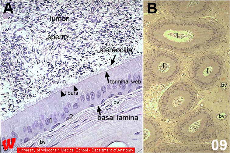
HA9 shows two LMs. B is a low magnification image of the epithelium of the epididymus (Cell Biology HA9). It shows multiple sections through a single coiled tube containing sperm in the lumen. A shows the same epithelium at higher magnification. At first glance the epithelium appears to be stratified columnar, but careful examination reveals that all cells touch the basal lamina. There are two rows of nuclei: one continuous row of nuclei in the center to lower third of the cells, and one staggered row of nuclei touching the base. The basal cells are not packed together tightly because they must leave room for processes of the apical cells to reach the basal lamina. At the apex, there are stereocilia - long microvilli that increase the surface area to absorb fluid and phagocytose defective sperm. The terminal web (faint line) and terminal bars (small dark dots) are visible at the apical regions of the cells. The complete name of this epithelium is pseudostratified columnar epithelium with stereocilia. Underneath the epithelium are a number of blood vessels (bv), since epithelium is avascular.