


HA6
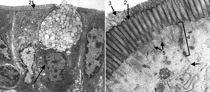
HA6 shows two EMs. At the left is a low magnfication EM of 4-5 cells similar to those in the LM in HA5. The second is a high magnification EM of part of the apical regions of two adjacent cells in the low mag EM. The two cell types present in the low mag EM are absorptive cells (enterocytes) which have abundant microvilli, and a goblet cell that has a cytoplasm full of vesicles of mucus. At the right, the high mag EM shows microvilli and the glycocalyx that is a component of their membranes. In the right third of this image is a junctional complex that is composed of a tight junction (TJ), an adhesion belt (AB) and a spot desmosome (SD). LCM is the lateral cell membrane. Parallel to the tight junction and adhesion belt, actin filaments can be seen extending from the cores of the microvilli to interact with the terminal web in the apical cytoplasm.