


HA5
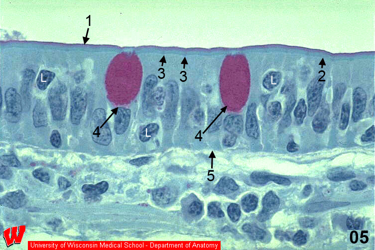
The epithelium covering the villi has a variable appearance. Some portions seem stratified due to the presence of lymphocytes (L) conducting immune surveillance, but the thinnest area is simple columnar epithelium, as shown in this high magnification LM image (HA5) which is perpendicular in orientation to HA4. Careful examination of this light micrograph reveals two stripes at the apical surface (top), one red stripe and one blue-green stripe. The red stripe is the glycocalyx, a layer of sugars linked to the cell membrane that stains red/pink with the sugar-specific PAS stain. The blue-green stripe is the microvillous border. At the base of the microvilli there is a thin, darker green line, the terminal web. Faint spots at the apical/lateral surfaces of the cells are terminal bars (junctional complexes). Scattered in the epithelium are red ovals, which represent vesicles in the cytoplasm of goblet cells that secrete mucus onto the surface. The basement membrane is a faint line above the lamina propria. The complete name of this epithelium is simple columnar epithelium with microvilli and goblet cells. This type of epithelium is found throughout the intestines. Note that the villus (HA4) is a gross anatomical structure easily seen at low magnification, whereas microvilli (HA5) cannot be seen even at high magnification in a light micrograph.