


HA18
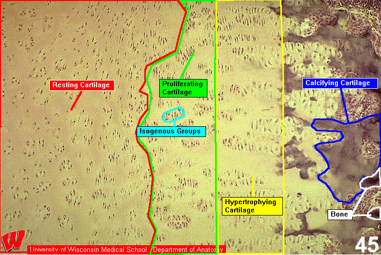
HA18 shows a higher magnification micrograph of the epiphyseal region of the distal phalanx. Although the zones blend into one another, it is possible to distinguish the typical appearances of the zones of resting/reserve cells (HA18 r, left), proliferating (p, center), hypertrophying (h), and calcifying (c, right) cartilage. b indicates the front edge of the ossification zone. The chondrocytes stack up like pancakes in the zone of proliferation and enlarge in the zone of hypertrophy.