
Home -> Unit 1 -> Nerve Tissue


HA17
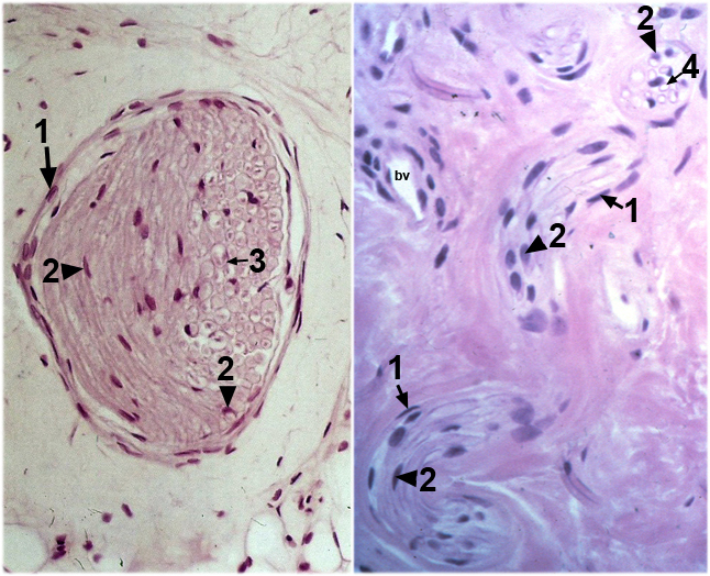
This image contains two examples of small peripheral nerve bundles, because these can be difficult to identify in sections. The LM on the left shows a PNB in transverse section. The motor axons, which run to peripheral muscle targets, cannot be distinguished morphologically from sensory axons, which run to the dorsal spinal cord from the periphery. Nuclei of perineurial cells wrap the PNB. The inner nuclei are Schwann cells if they are associated with axons and endothelial cells if they are associated with blood vessels. The right panel shows a PNB in cross section at the top right of the image and in two longitudinal sections in the middle and lower parts of the image. The PNB moves in and out of the plane of section because of its tortuous structure. Schwann cell nuclei and myelin are visible in both planes of section. In a few places a clear axon cytoplasm can be seen wrapped by myelin. bv is a small blood vessel.