
Home -> Unit 1 -> Nerve Tissue


HA15
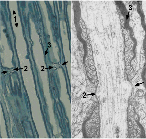
The LM image on the left shows several axons in longitudinal section in a PNB. The pale staining central regions are the axon cytoplasms. At the arrows, the myelin wrapping of one Schwann cell ends and that of the next one along the axon begins. This tiny gap in myelination is called a node of Ranvier (after the scientist who first described them). The node permits saltatory (jumping, rapid) conduction of the action potential (AP). The EM image on the right shows a node of Ranvier at very high magnification. Ruffling of the Schwann cell membrane occurs on either side of the node. This open area of axon cell membrane is full of ion channels that maintain the flow of the AP. What cytoskeletal elements are present in the axon in the EM image?