
Home -> Unit 1 -> Nerve Tissue


HA9
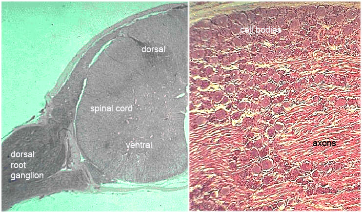
The very low magnification image at the left shows one of two ganglia (cluster of neuron cell bodies outside the CNS) that are located at the sides of the spinal cord. These spinal/dorsal root ganglia are made up of groups of unipolar neurons alternating with bundles of axons. These sensory axons enter the spinal cord at the dorsal root, which is the basis of the name. (The ventral horn neurons are not prominent in this sample.) The low mag LM image of the spinal ganglion at the right shows this arrangement of round neuron cell bodies with filamentous axons. This section is shown at higher magnification in HA10.