
Home -> Unit 1 -> Nerve Tissue


HA6
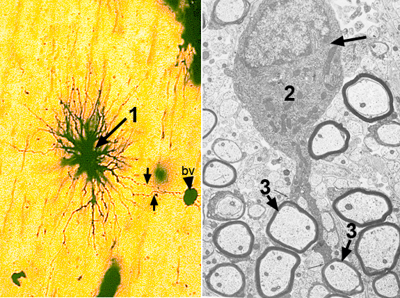
The image on the left is a LM of an astrocyte. In this histological preparation all of the processes of a single astrocyte are filled with black stain. Other cells in the region are unstained. At the bottom and right, blood vessels have also taken up the black stain. One of the functions of the astrocyte is to make contact with neurons of the CNS to help maintain their ionic micro-environments. Excess ions are moved through astrocyte processes to blood vessels into which they diffuse.
The image on the right is an EM of an oligodendrocyte. Its function is to myelinate axons in the CNS. In this image trace two oligo processes from the cell to two myelinated axons.