
Home -> Unit 1 -> Nerve Tissue


HA3
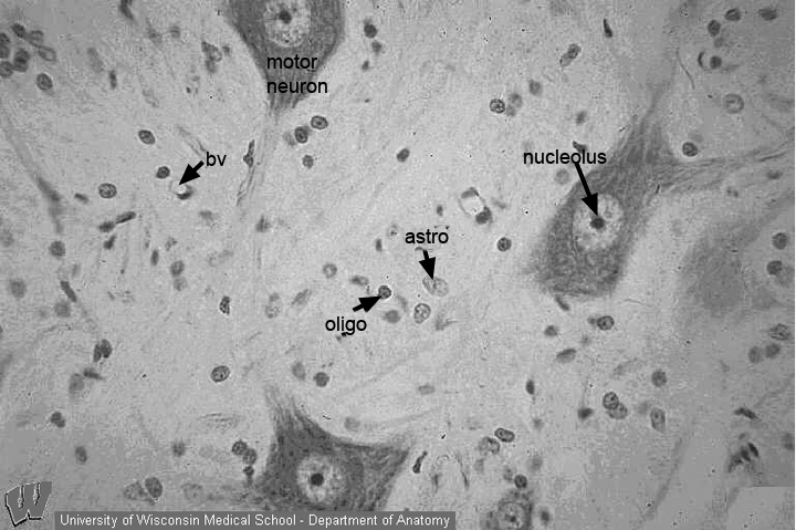
At high magnification in the same area as that shown in HA2, cytoplasmic details and nucleoli can be seen. The cytoplasm of these large neurons is packed with both bound (RER) and free ribosomes. These cells require large numbers of proteins, such as the cytoskeleton (Cell Biology HA1), cell membrane proteins (ion channels) and polypeptides (neurotransmitters for exocytosis/cell signaling), for normal function. These proteins are made in the cell body and distributed in axons that can be 3 or more feet in length.
The oligodendroglial cells, which myelinate axons in the CNS, have small, round, comparatively heterochromatic nuclei. The nuclei of astrocytes (HA5, A) are larger and more euchromatic than the nuclei of oligodendrocytes. Endothelial nuclei of small blood vessels (bv) are most easily identified in cross sections. The nuclei are small, heterochromatic, and appear flattened at the edge of the vessel lumen.
The presentation of this image in grayscale illustrates the principle that colors are not required for you to be able to identify cell structures in light micrographs.