
Home -> Unit 1 -> Nerve Tissue


HA2
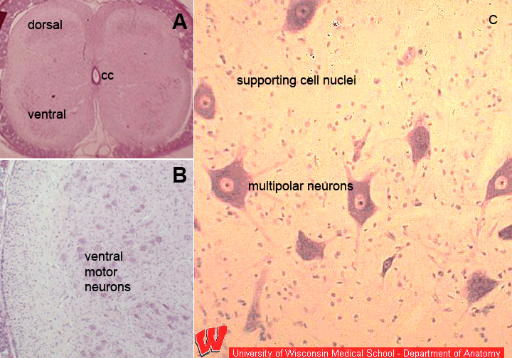
This montage (composite image) shows a very low magnification image of a spinal cord at the upper left (A). The boxed area containing motor neurons of the ventral horn is shown at low magnification at the lower left (B). At the right is a medium magnification image showing a subset of these cells (C). These are multipolar neurons (HA7) with large nuclei, and prominent nucleoli.
Due to the stain used, there is a poorly staining region with little apparent structure that lies in between the large neuron cell bodies. This region, which is called the neuropil, when examined with the electron microscope (HA5), is completely packed with dendrites, axons, capillaries, and glial cells, the nuclei of which are detected in this image as small dots. cc is the central canal in A.
This montage illustrates what you would see if your were looking at the section in A with a microscope objective that is low magnification (4x). As you change objectives to higher magnification, such as 10x (B), you focus in on the ventral horn (lower left) and then change to 20x for the image at the right (C).