
Home -> Unit 1 -> Nerve Tissue


HA1
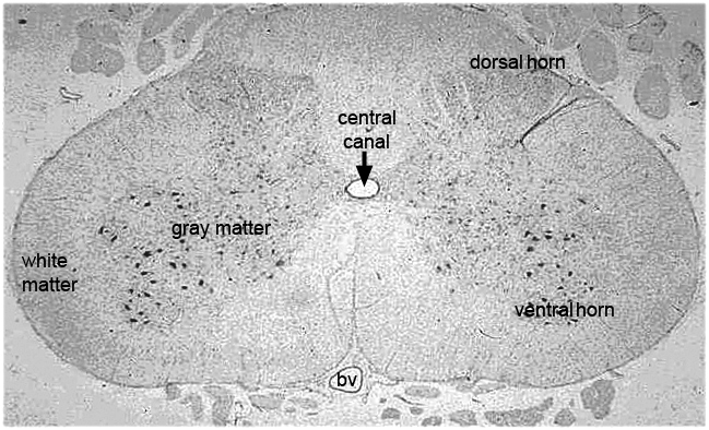
HA1 is a very low magnification image of a transverse section of an entire spinal cord. The overall arrangement of the gray matter (neuron cell bodies) in a section like this one has been compared to the shape of a butterfly with its wings spread. It stains as darker gray in this micrograph. The large dots are the cell bodies of the ventral horn. These very large neurons send out motor axons to the skeletal musculature. The axons of sensory neurons enter the region of the spinal cord called the dorsal horn. White matter, which contains glia but not neuron cell bodies, surrounds the gray matter. The central canal is filled with cerebrospinal fluid. Dorsal (toward the back) is at the top of this image, and ventral (toward the abdomen) is at the base. A large blood vessel is located ventrally.