


HA18
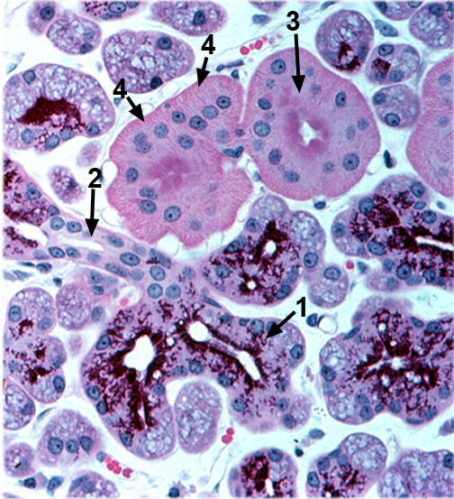
HA18 shows serous acini that are part of a salivary gland called the parotid. In this uncommon histological stain, the serous cells have densely stained vesicles of secretory material in their apical cytoplasms. The ducts that connect them to the oral cavity are of two types. The duct that leads out of each acinus is made of simple squamous to cuboidal epithelium. The fluid flows into a striated duct which contains simple columnar epithelia, in which the nuclei are central rather than basal. This epithelium actively modifies the ion content of the saliva by pumping ions through transmembrane channels that are located in basal infoldings of the cells. These appear as faint lines at the bottom of striated duct cells. A blood vessel runs at the base of the striated ducts to facilitate the ion exchange. L is the lumen of an adenomere.