


HA11
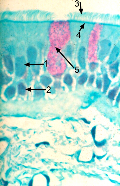
The trachea is also lined with pseudostratified columnar epithelium, with two layers of nuclei, but it has different apical modifications that are necessary for its function. In this medium magnification light micrograph, at the apex of the epithelium, you can see individual structures projecting into the lumen. These are cilia. Because they are thicker and longer than microvilli, they are visible in light micrographs. In the cytoplasm beneath the cilia there is a dark line where the basal bodies of the cilia are embedded in the terminal web. Cilia contain microtubules rather than actin filaments, and move in unison toward the oral cavity so that mucus and trapped particulates can be removed from the respiratory tract.
Scattered among the ciliated cells there are magenta stained goblet cells. The complete name for this epithelium, with its modifications, is pseudostratified columnar epithelium with cilia and goblet cells. Because it is found only in the lungs, it is often called respiratory epithelium. In the underlying lamina propria (loose connective tissue layer), there are blood vessels (bv).