


HA1: Human ovary H&E
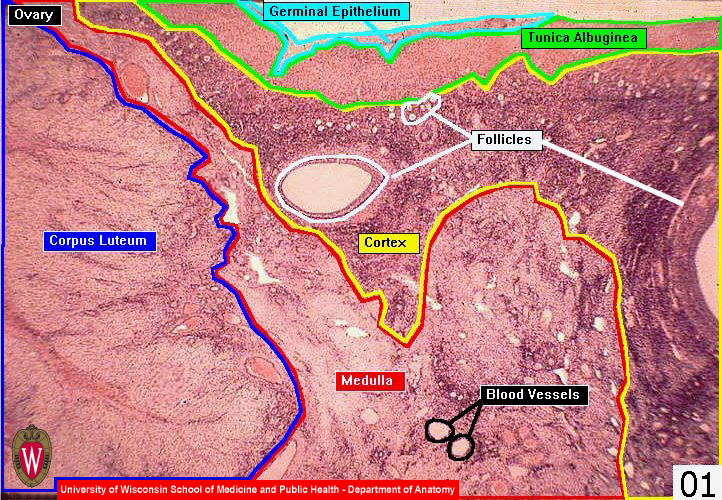
At the top of this low magnification image of an ovary (HA1), you can see the germinal epithelium (asterisk) on the surface of the ovary and the underlying connective tissue layer, the tunica albuginea (ta). The cortex (C) is composed of follicles embedded in a connective tissue stroma, which is extremely cellular. Cells from the stroma are recruited to form the thecas, interstitial glands, and paralutein cells described in subsequent slides. Beneath the cortex is the more vascular medulla (M). As an endocrine organ, the ovary has a rich vascular supply. A portion of a corpus luteum (cl) is present in this micrograph. It appears to be in the medulla due to the plane of section. Follicles (f) in a variety of sizes are present in the image.