


HA4: Kidney, human H&E
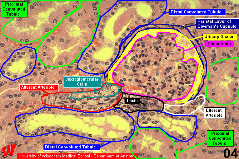
In the renal corpuscle, you can see the urinary space (HA4, u) between the parietal and visceral layers of Bowman’s capsule. The parietal layer of the capsule is a simple squamous epithelium with flattened, heterochromatic nuclei. The visceral layer is a unique epithelium made up of podocytes that form part of the urinary filter. Podocyte nuclei can be distinguished because they are large and protrude into the urinary space. In light micrographs, it is difficult to distinguish between endothelial and mesangial cells within the corpuscles, but the distinction is more obvious in electron micrographs (see the Urinary System EMs pdf)
An afferent (HA4, a) and a small section of an efferent (HA4, e) arteriole can be seen at the vascular pole of the renal corpuscle. The afferent arteriole is distinctive because it has juxtaglomerular cells (HA4, JG) in its tunica media. JG cells are modified smooth muscle cells that secrete renin. The JG cells are one of three components of the juxtaglomerular apparatus. The other two components are the macula densa (HA5 m) and the lacis, which is composed of a compact mass of extraglomerular mesangial cells (HA4, m) that migrate into the renal corpuscle to form phagocytic cells to remove debris from the urinary filter. Because of their spatial relationships, it is rare to find all three elements of the JG apparatus in a single section.
The cortical tissue surrounding the renal corpuscles is packed mostly with sections of proximal convoluted tubules (HA4, P) (PCT) and distal convoluted tubules (HA4, D) (DCT). Although their names sound similar, the PCT and DCT have very different morphologies. The PCT cells have such extensive microvilli that their apical surfaces are fuzzy and the lumen of the tubule is often obscured. The cytoplasm of DCT cells is paler and the apical surface is more distinct because there are many fewer microvilli on these cells. Cells in both the PCT and DCT have extensive lateral interdigitations, so the intercellular boundaries are difficult to see in both structures.