


HA1: Kidney, human H&E
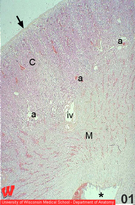
This low magnification image of a human kidney (HA1) shows the connective tissue capsule (arrow), the cortex (C) and medulla (M) with a minor calyx (asterisk) at the bottom. A large interlobar vessel (iv, HA1) branches to form arcuate vessels (HA1, 2 and 3, a) that travel along the boundary between the cortex and medulla.