
Home -> Unit 1 -> Cell Biology


HA2
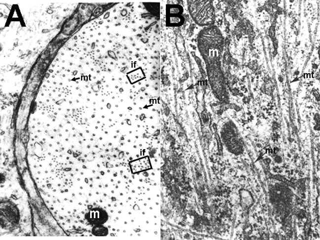
The two panels, A and B, in this image are high magnification electron micrographs (EMs) that allow us to see the structure of the cytoskeleton. A is in transverse (cross) section and shows microtubules and intermediate filaments (if; neurofilaments) in a neuron. B is in longitudinal section and shows microtubules running from the top to the bottom of the image among other organelles such as mitochondria (m).
Within an image, when you see the same number used more than once, you are looking at the same cell structures/organelles.