


HA16: Esophagus, cat, H&E
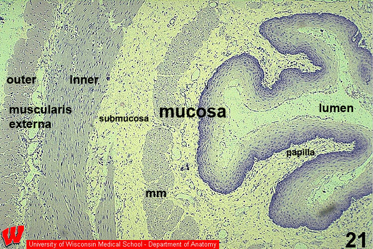
The esophagus in HA16 shows the basic layered structure of the digestive tract: mucosa, submucosa, and muscularis externa. The fibrosa that surrounds this organ is not evident in this micrograph. The basal region of the stratified squamous mucosal epithelium is indented with connective tissue papillae of the lamina propria. The muscularis mucosae (mm; cut in cross-section) is thick and separates the inner lamina propria (loose CT) from the outer submucosa (dense irregular CT). The inner circular layer of the muscularis externa is mostly smooth muscle with a few skeletal myofibers, while the outer longitudinal layer is mostly skeletal muscle (in cross-section).