


HA13: Intrapulmonary bronchi
As the conducting portion of the respiratory system enters the lungs there are two main changes in the supporting tissue composition that occur gradually with each branching. (1) The hyaline cartilages in the wall are arranged with decreasing regularity. In the extrapulmonary (primary) bronchi, there are cartilage rings that wind around the tube, often with a helical configuration. In the intrapulmonary bronchi, the cartilages have the form of irregular plates that overlap with each other along the length of the tube maintaining firm support for the airway. (2) Bundles of smooth muscle and elastin become more prominent in the lamina propria.
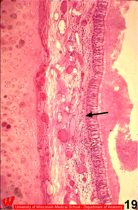
There are two sections of an intrapulmonary bronchus shown in HA13&14. One was stained with H&E (HA13) to show general cellular detail, and the other was stained for elastin (HA14) to highlight its abundance in the respiratory CT. In HA13,the blanket of mucus is in the lumen above the cilia. The thin layer immediately surrounding the cilia is a serous fluid that stains more lightly. This serous fluid allows the cilia to beat in unison to move the mucus toward the epiglottis. The basement membrane in the conducting portion is thick (arrow, HA13)