


HA10: Trachea and esophagus, monkey
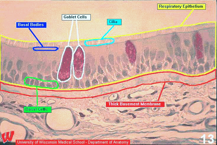
At higher magnification, HA10 shows the respiratory epithelium lining the trachea. Note the clearly defined cilia and the line in the apical cytoplasm (arrows) delineating the basal bodies in the terminal web. The mucopolysaccharides in the cytoplasm of the goblet cells stain red. Basal cells are stem cells. Two other cell types, brush cells and granule cells, are also present, but are not discernible from other cell types with conventional histological stains. Below the epithelium are blood vessels in the loose CT. The epithelium is kept moist not only by goblet cells, but also by serous and mucous glands in the lamina propria (HA7).