
Home -> Unit 4 -> Cardiovascular 2


HA18: Diad EM
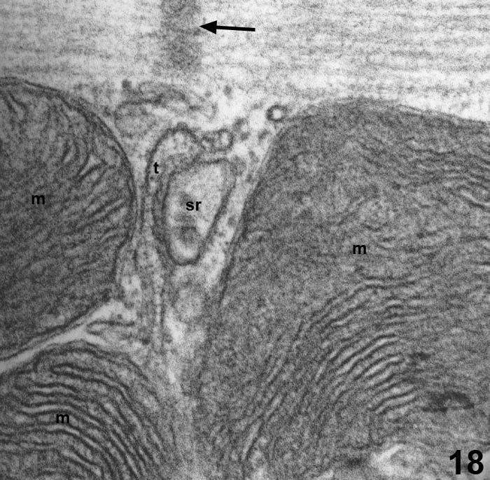
This high magnification electron micrograph shows a T-tubule (t) and a cistern of sarcoplasmic reticulum (sr) at the Z line (arrow) of a cardiac myocyte. On either side of the diad are mitochondria (m).