
Home -> Unit 4 -> Cardiovascular 2


HA17: Intercalated disk EM
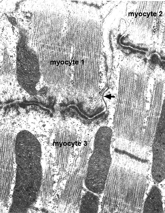
This electron micrograph shows three cardiac myocytes connected by intercalated disks. Myocyte 1 and 3 are electrically coupled by a gap junction (arrow). The dark staining lines are spot desmosomes and actin filaments inserting on the sarcolemma, which is known as a fascia adherens. There are mitochondria in between the myofibrils.