
Home -> Unit 4 -> Cardiovascular 2


HA16: Purkinje fibers
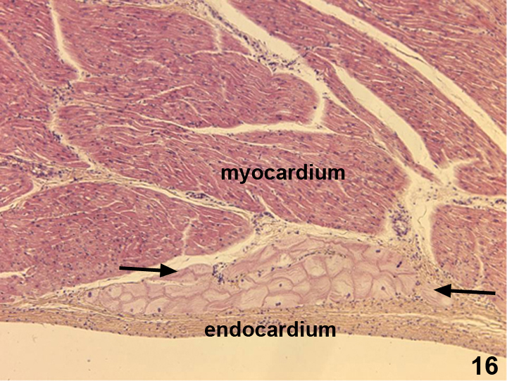
This image shows modified cardiac myocytes called Purkinje fibers (between the arrows) that run in the subendocardial CT space. These cells carry the action potential from the pacemaker to the tip or apex of the ventricle to initiate contraction.