
Home -> Unit 4 -> Cardiovascular 2


HA7: Left ventricular wall
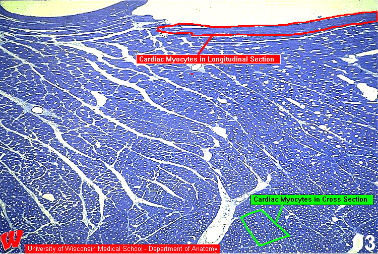
Most of the myocytes in these images are seen in cross section, but the orientation of the myocyte fascicles gradually changes across the myocardium so that near the endocardium (top) they are in longitudinal section (HA7). This appearance is due to the way in which the fascicles wrap around the ventricular lumens. Arrows point to perimysium.