
Home -> Unit 4 -> Cardiovascular 2


HA5: Left ventricular wall
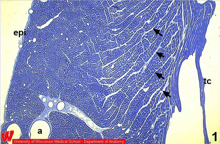
In this low magnification micrograph (HA5), it is evident that almost the entire thickness of the wall of the ventricle is made of myocardium. On the outer surface (left), the thin epicardium (epi), with 3 vessels running in its underlying CT layer, is visible. Branches of the coronary artery (a) and cardiac vein can be seen within the myocardium (HA5, bottom left). This and the following images (HA6-9) are from a heart perfused and fixed under pressure, so all the blood vessels are abnormally large in diameter. The inner surface of the ventricle is irregular because of folds in the muscle called trabeculae carneae (tc). The endocardium is so thin that it can’t be seen at this low magnification. The myocardium contains thin connective tissue septa (arrows) that define fascicles of myocytes (HA5&7).