
Home -> Unit 4 -> Cardiovascular 2


HA3: Papillary muscle
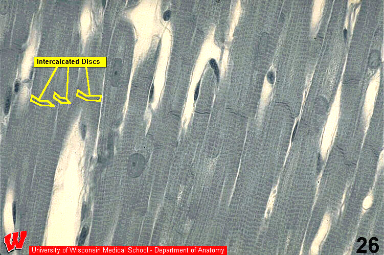
The diagnostic feature of cardiac muscle in longitudinal section is the intercalated disk. These disks are at the junctions between adjacent myocytes. At lower magnification, the intercalated disks can look like a flight of stairs (HA3). The connective tissue sheathes that organize skeletal muscle are present in cardiac muscle, but they are much thinner because of the intercalated disks. The capillaries (HA3 c) and their underlying loose CT are the only component of the endomysium that is visible in these images.