
Home -> Unit 4 -> Cardiovascular 2


HA1: Papillary muscle
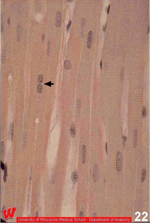
The first four micrographs in this lab exercise aresections of cardiac papillary muscle. The first is stained with H&E (HA1), the others with iron hematoxylin (HA2-4). Several features in these images make it possible for you to identify this striated muscle as cardiac muscle. The nuclei are large, euchromatic, and centrally placed in the myocytes (HA1-2). Most myocytes have a single nucleus, but some are binucleated (HA1 arrow). You will not see more than two nuclei in one myocyte.