


HA4
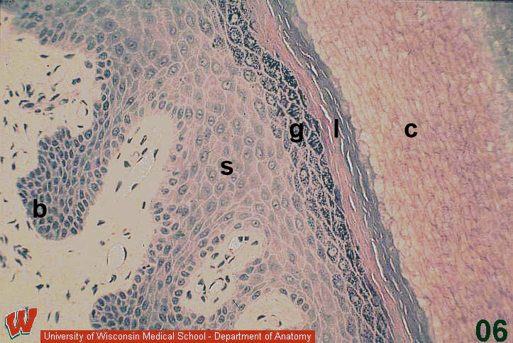
In this micrograph (HA4) of thick skin, you can distinguish five layers of the epidermis (but the outermost portion of the stratum corneum is not visible in this image). Starting at the basement membrane, cells in the stratum basale (b) vary between columnar and cuboidal. This is the layer where stem cells divide to populate the other layers. Cells in the stratum spinosum (s) have a slightly more flattened shape and appear to be covered with spines because of the spot desmosomes that bind them together. Cells in the stratum granulosum (g) are even more flattened and have prominent dark granules in their cytoplasm. Cells in the stratum lucidum (l) are dead. Note that they lack nuclei. Cells in the stratum corneum (c) are dead and flattened into squames. We do not know why the staining is variable in the stratum corneum.