


HA15
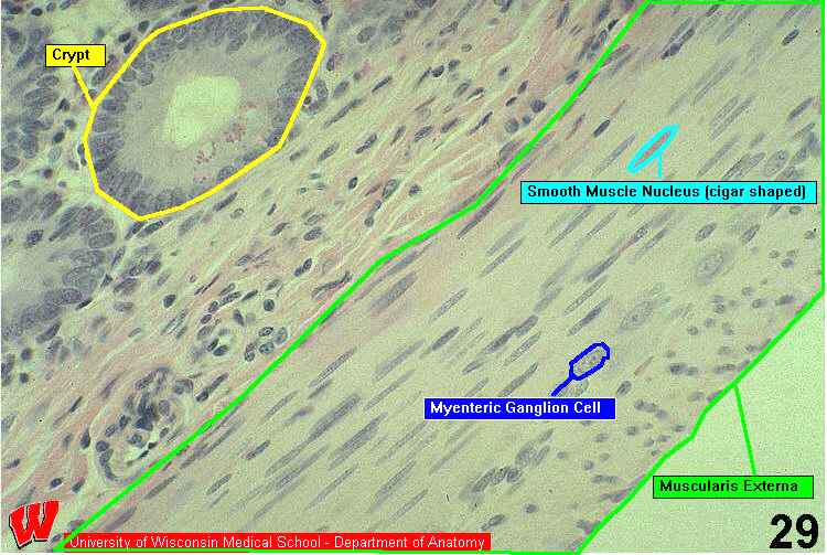
HA15 is a higher magnification image in which you can distinguish the inner and outer layers of the muscularis externa. The outer, longitudinal layer of the muscularis externa shows smooth muscle myocytes cut in cross section (HA15). Notice that the myocyte margins cannot be resolved easily; however, it is possible to see the cigar-shaped nuclei (HA15). Because the myocytes overlap each other, the nuclear cross sections vary considerably in their apparent diameters. This is a diagnostic structural feature of cross sections of smooth muscle. The inner, circular layer shows longitudinal sections of myocytes.
Between these two layers of smooth muscle there are groups of neuron cell bodies (ganglia) that innervate the two layers of smooth muscle (HA15). Look for the larger oval nuclei with pink nucleoli at the right center. This is a myenteric ganglion, which is composed of postganglionic parasympathetic neurons and their processes. Similar ganglia are found just inside the inner layer of smooth muscle but are not shown here. These cell bodies and their processes compose the submucosal ganglion.