


HA17
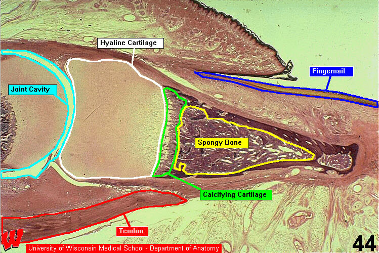
The low magnification light micrograph in HA17 shows the joint cavity (c) between the medial and distal phalanx (distal bone) of the finger. The dorsal epidermis is at the top and the fingernail is at the right. Secondary ossification centers have not yet formed in the epiphyses. A primary ossification center has formed in the distal phalanx and has a rich red marrow. There are two tendons, which attach to muscles (not shown) lower in the finger.