


HA 38
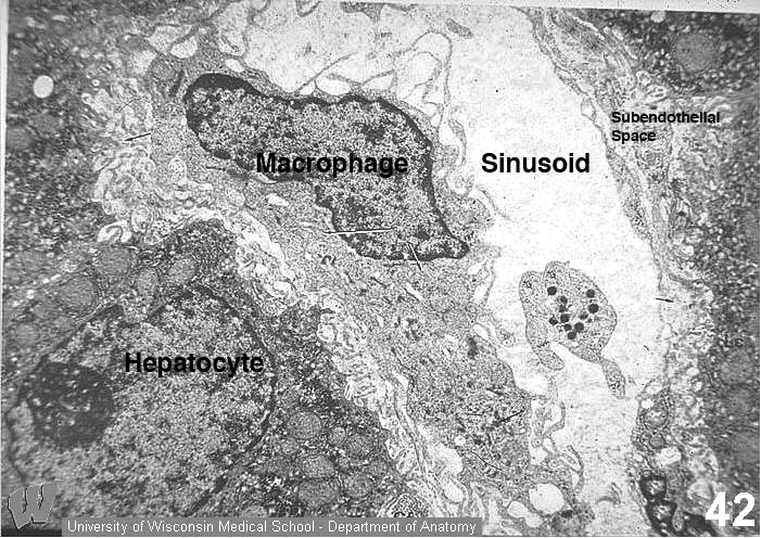
In this micrograph, a Kupffer cell (Kc) forms part of the wall of a sinusoid (Slu). Notice the numerous ruffles on the macrophage that reflect its phagocytic function. Processes of this cell project into the subendothelial space of Disse (Ds). A platelet (Pl) is in the sinusoid. Ed=endothelial cell, Ly=lysosome, m=mitochondrion, Mv=microvilli, Cf=collagen (reticular) fibers, Go=Golgi, N=nucleus, Nu=nucleolus.