


HA21
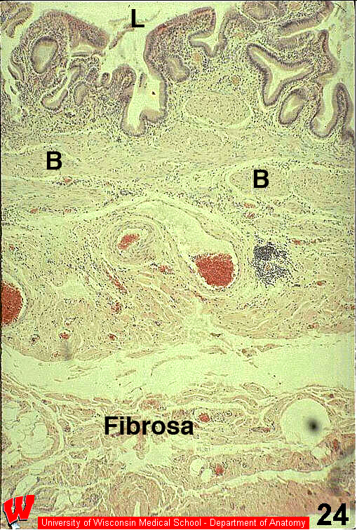
HA21 shows the layers of the gall bladder wall. At the top is the highly folded mucosa consisting of the simple columnar epithelium with microvilli and the underlying loose connective tissue of the lamina propria (L indicates the lumen). The next layer below is the muscularis externa consisting of large, irregularly oriented bundles of smooth muscle (HA21, B). The third layer is a fibrosa, since mesothelium and the peritoneal cavity are not evident. This section was taken from the thicker wall of the gall bladder where it is attached to the liver. http://www.medicalart-dank.com/promotion/33042LG.jpg Large blood vessels, adipose tissue and much dense irregular connective tissue are present within the fibrosa.