


HA1
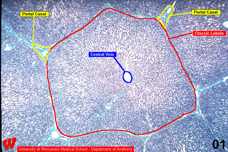
This image HA1 is a low magnification light micrograph that shows the classic hexagonal liver lobule very well. These lobules can vary from four-sided to eight-sided. Note the central vein which is close to the middle of the lobule (C in HA1) and portal canals at its corners (P in HA1).