
Home -> Unit 1 -> Histopathology


HA4
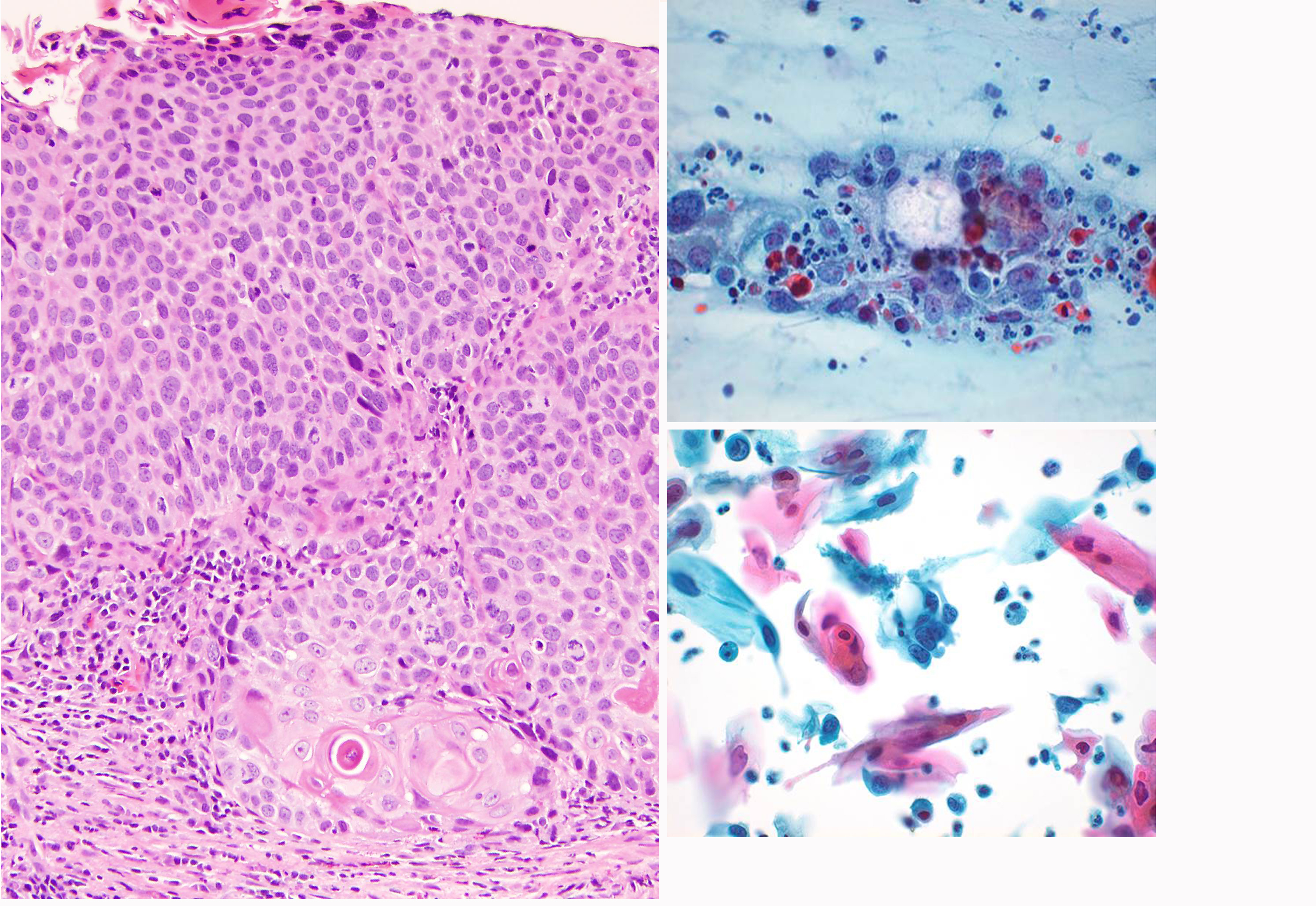
This micrograph shows a biopsy/resection specimen on the left and pap smears on the right. The biopsy has high grade dysplasia, with loss of polarity of the epithelium (larger nuclei and high N:C ratio cells from top to bottom), with mitotic figures (black arrows) in the midst of the epithlium. The nuclei are irregular in shape and have small nucleoli. At the bottom is a focus of invasive carcinoma (black circle), which has broken through the basement membrane and incited an inflammatory cell response in the surrounding stroma. In this case, the cells have become keratinizing (the dark pink) but this does not have to be a feature of invasive squamous cell carcinoma. The pap smear shows high grade dysplasia in the cells (as in HA3) but also shows necrotic debris (blue arrows) and inflammatory cells (neutrophils, red arrows) which strongly suggests that the high grade dysplasia has progressed to an invasive cancer. (In house micrographs)