
Home -> Unit 1 -> Histopathology


HA3
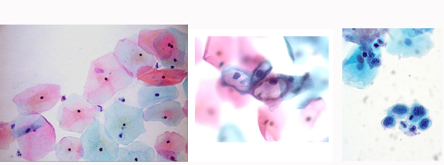
These micrographs are taken from smears of cervical scrapings that are stained with the Papinicolaou (“Pap”) stain. On the left are normal superficial squamous cells (the top layer of cells from a cervix that looks like HA1 and the right hand side of HA2). Note the very small nuclear:cytoplasmic (N:C) ratio, with small pyknotic nuclei that have smooth nuclear contours. The red vs. blue staining of the cytoplasmis not important. (internal photo)
The center photo represents low grade dysplasia (also called low grade squamous intraepithelial neoplasm [LGSIL] or cervical intraepithelial neoplasm-1 [CIN-1]). Note that there are some normal cells in the background, but the cells in the center of the photo have somewhat larger nuclei that have grooves or notches in their nuclear membrane, more open (pale) chromatin, and what seem to be cleared out areas around the nuclei (arrows). These changes are due to human papilloma virus (HPV) infection and these cells are called koilocytes (from the Greek, koilos [hollow] and kytos [cell]). One could imagine such cells being scraped off the left side of the cervix in HA2. (internal photo)
The right hand photo shows high grade dysplasia (HGSIL or CIN2-3), particularly in the cell cluster at the lower right, with high N:C ratios, irregular nuclear contours and more coarse chromatin. (© Ed Uthman, M.D., CC-BY, http://www.flickr.com/photos/euthman/sets/72057594114099781/with/284183160/