
Home -> Unit 1 -> Histopathology


HA2
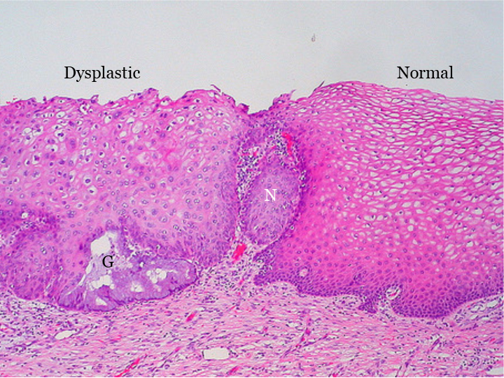
This photomicrograph shows normal (right) and dysplastic (left) squamous epithelium in the cervix. Note the progressive decrease in nuclear size in the normal area as you approach the surface. Although that is also seen in the dysplastic epithelium, on average, it is also possible to find larger nuclei near the lumen and generally much further “up” the epithelium. Nucleoli are evident in some of these cells midway up the epithelium. There is a small focus of glandular epithlium (G) which appears benign – the dysplastic squamous epithelium likely has “crawled” over this gland. The small nest of cells in the center (N) is not invasive cancer but rather a tongue of dysplastic epithelium that connects up to the surface out of the plane of this slice through the tissue. This photo demonstrates how dysplasia tends to be a focal process, the result of individual cells acquiring genetic lesions and giving rise to colonies (clones) of dysplastic progeny. (© Ed Uthman, M.D., CC-BY, http://www.flickr.com/photos/euthman/sets/72057594114099781/with/284183160/ )