
Home -> Unit 1 -> Nerve Tissue


HA24
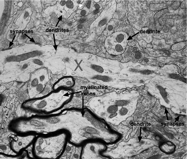
This high magnification EM of the CNS shows a dendrite (X) traversing the middle of the image. It's cytoplasm contains mitochondria and cytoskeletal elements. The darkly outlined structures are myelinated axons. Dendrites can have many hundreds of axodendritic synapses depending on their length and amount of branching. Close examination of densely staining regions of the dendritic membrane reveal that these are synaptic densities and that many synaptic vesicles containing neurotransmitter rest next to the synapses. A careful examination of this entire micrograph will allow you to see many dendrites in different planes of section. Each is surrounded by axon termini although the synaptic densities are often not in this plane of section.