
Home -> Unit 1 -> Cell Biology


HA23
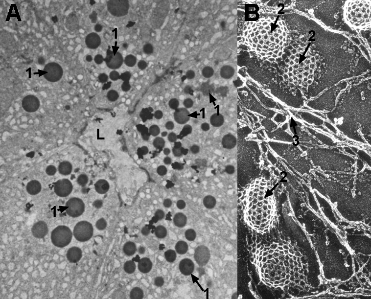
The EM image in A shows vesicles in the apical cytoplasms of an exocrine gland. This is an example of regulated exocytosis. The vesicles, which stain black, contain proteins such as enzymes for release when a hormonal or neural signal is received. L is the lumen of the adenomere. B is a scanning EM image of the inner surface of a cell membrane. The cage-like structures are clathrin-coated vesicles or coated pits for endocytosis. The membrane is stabilized in part by the cytoskeleton.