
Home -> Unit 2 -> Cardiovascular I


HA19
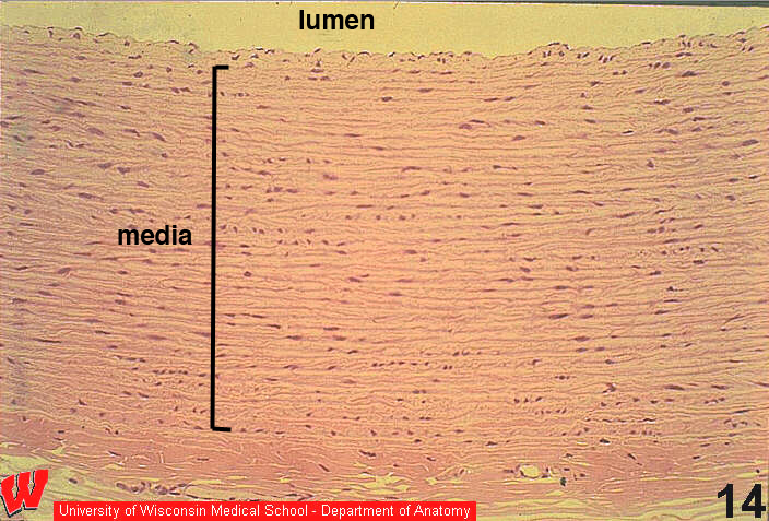
This image shows a cross-section of the wall of an aorta and a portion of the vessel lumen (top). Identify the layers in the wall of the aorta. The tunica intima consists of endothelium and a thin layer of connective tissue. Its thickness is not obvious, but blends into the tunica media, which occupies most of the image. Because this is an elastic artery, it has multiple (30-35) elastic laminae that are secreted by the intervening smooth muscle cells. The outermost layer, the tunica adventitia, is a layer of connective tissue that contains nerves and the vasa vasorum that supply the tunica media.