
Home -> Unit 2 -> Connective Tissue


HA1
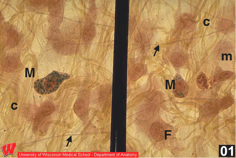
The image in HA1 is not a section; it was made by fixing and staining a piece of mesentery, then spreading a small piece onto a slide. A mesentery is a thin layer of loose connective tissue sandwiched between two layers of simple squamous epithelium. Anatomically it is the support for the intestines in the abdominal cavity. Its loose connective tissue illustrates three of the four main cell types and the collagen and elastic fibers that are typical of connective tissues.
The simple squamous epithelium lining both sides of a mesentery is called mesothelium. These cells are so thin that only their flattened, pale staining nuclei are discernible (mesothelial nuclei, HA1, m). In the connective tissue, collagen fibers (HA1 c) appear as diffuse, wavy, yellow bundles in the matrix. Collagen I predominates in this loose connective tissue. Elastic fibers (HA1, arrows) appear as sharp, straight lines with occasional branches. Note the relative numbers of collagen and elastic fibers.
The predominant cell in CT is the fibroblast (HA1, F). This cell has a round to oval nucleus, a prominent nucleolus, and densely staining cytoplasm. Fibroblasts are active cells that maintain the connective tissue matrix. The second major connective tissue cell type is the mast cell (HA1, M). Mast cells are small, densely staining, and round or oval. The nuclei are usually obscured by many cytoplasmic granules that stain green in these micrographs. These granules contain several important compounds including heparin and histamine.