
Home -> Unit 1 -> Cell Biology


HA7
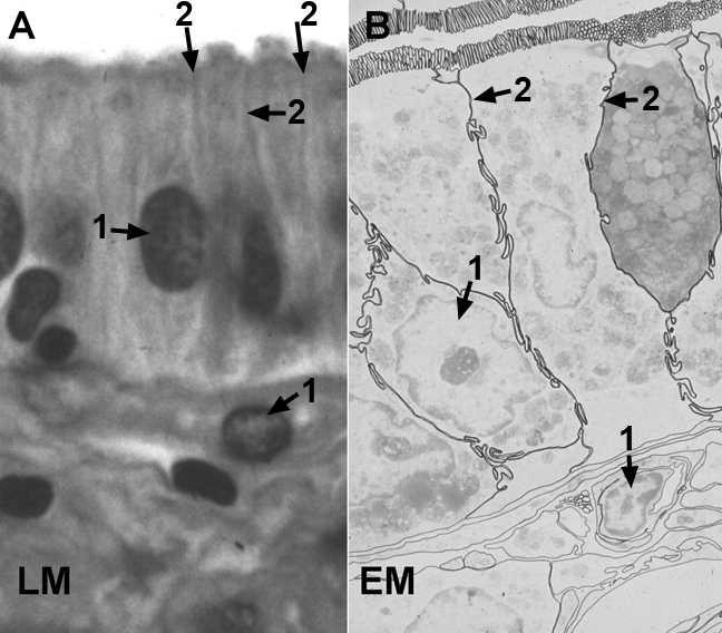
HA7 highlights the appearance of cell membranes in a light micrograph (A). Even in high magnification LM images, cell membranes can be somewhat difficult to see. In this image the lateral membranes appear as dark fuzzy lines between cells. This section was stained with a histological dye, but presentation here as a grayscale image improves the contrast of the membranes. In B, a low magnification EM image, the lines of the apical, lateral and basal cell membranes have been traced and highlighted. The normally prominent cell organelles such as the nucleus have reduced contrast to permit you to see the folding of the membranes between adjacent cells (lateral interdigitations), the large number of microvilli on the apical membrane and the comparatively flat basal membrane. Even at low magnification, EMs significantly improve the resolution of fine structures in cells.