
Home -> Unit 2 -> Cardiovascular I


HA22
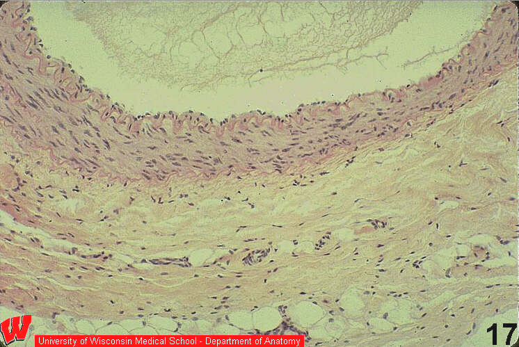
The layers of the arterial wall are sharply defined (HA22) in this light micrograph. There is a tunica intima delimited by an internal elastic lamina (pink) that is convoluted because of vasoconstriction. The tunica media at its narrowest point has more than 5 layers of smooth muscle cells (count the nuclei ). The tunica media ends with the external elastic lamina. Beyond this, the tunica adventitia is about one-half the thickness of the tunica media. The tunica media is always thicker than the tunica adventitia in arteries and arterioles. This sample was prepared from a cadaver. The artery lumen contains clotted serum, and most of the clotted blood is found in the vein, where it pooled.