(HA1-12): Kidney, human H&E
|
|
|
|
|
|
|
|
|
|
|
|
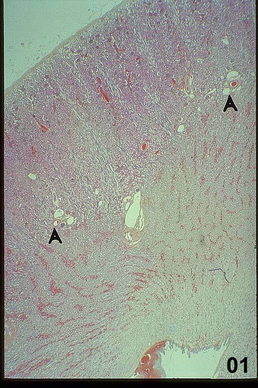
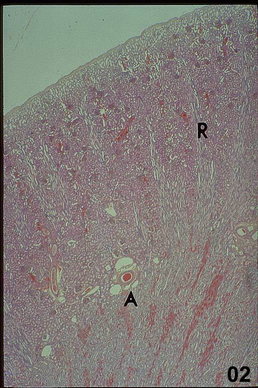
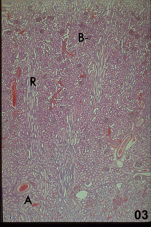
This low magnification image of a human kidney (HA1) shows the capsule and the cortex (top) and medulla (below) with a minor calyx at the bottom. A large interlobar vessel (center HA1) branches to form arcuate vessels (HA1, 2 and 3, A) that travel along the boundary between the cortex and medulla. Interlobular vessels (HA1-3) branch from the arcuates and run in the cortex. At higher magnification, medullary rays (HA2 and 3, R), which are several parallel clear lumens, are projections of the medulla that extend into the cortex. A medullary ray and its associated cortex form a renal lobule.
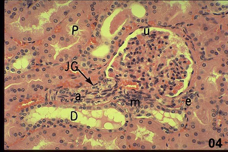
It is easy to identify the cortex because it contains all of the renal corpuscles (HA3-B). They look like small, dark knots, and each contains a capillary bed called the glomerulus and its surrounding Bowman’s capsule. In the right plane section of a renal corpuscle, you will see the urinary space (HA4, u) between the parietal and visceral layers of Bowman’s capsule. This is where the filtrate first accumulates. The parietal layer of the capsule is a simple squamous epithelium. The visceral layer is formed by podocytes that form part of the urinary filter. It is difficult to distinguish among podocytes, endothelial and mesangial cells within the corpuscles, but sometimes podocyte nuclei can be distinguished because they are larger and protrude into the urinary space. However, the distinction is more obvious in electron micrographs that you will look at later in this exercise.
In the right plane of section, afferent (HA4, a) and efferent (HA4, e) arterioles can both be seen at the vascular pole of the renal corpuscle. The afferent arteriole is distinctive because it has juxtaglomerular cells (HA4, JG) in its tunica media. JG cells are modified smooth muscle cells that secrete renin. In this image, the renin granules were extracted during preparation so the JG cell cytoplasm is clear. The JG cells are one of three components of the juxtaglomerular apparatus. The other two components are the macula densa and the lacis (HA4, m). The macula densa (HA5L) forms at the junction of the ascending thick limb and distal convoluted tubule where the tubule contacts its renal corpuscle. Macula densa means “dense spot” and this name makes sense when you see its tightly packed nuclei. In addition they show reverse polarity, so their nuclei are found apically, close to the DCT. They secrete from their basal surfaces to send paracrine signals to the JG cells to stimulate renin secretion.
The third element of the JG apparatus is the lacis. It is composed of a compact mass of extraglomerular mesangial cells (HA4, m) that occasionally migrate into the renal corpuscle to form intraglomerular mesangial cells, phagocytic cells that remove debris from the urinary filter. Because of their spatial relationships, it is rare to find all three elements of the JG apparatus in a single section.
The cortical tissue surrounding the renal corpuscles is packed mostly with sections of proximal convoluted tubules (HA4, P) (PCT) but also with distal convoluted tubules (HA4, D) (DCT). Although their names sound similar, the PCT and DCT have very different morphologies. The PCT cells have pink cytoplasm and such extensive microvilli that their apical surfaces are fuzzy and the lumen of the tubule is often obscured. The cytoplasm of DCT cells is paler and the apical surface is more distinct because there are many fewer microvilli on these cells. In addition, the nuclei are spaced irregularly in the DCT. Cells in both the PCT and DCT have extensive lateral interdigitations so the intercellular boundaries are difficult to see in both structures.
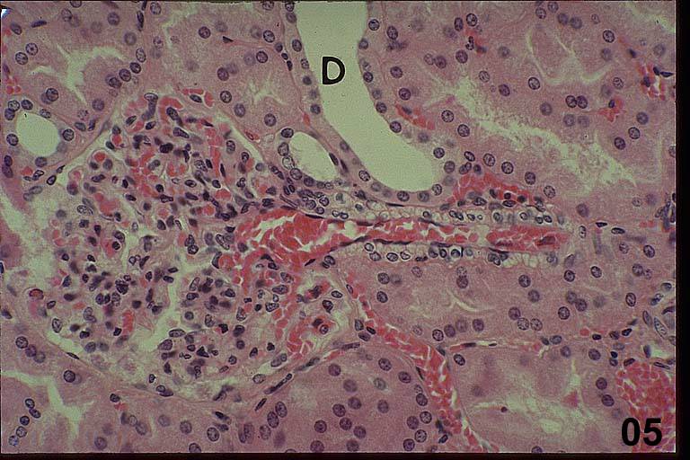
Occasionally, it is possible to find a section of a corpuscle that shows both its vascular and urinary poles (HA5). This image also shows good examples of an afferent arteriole, a macula densa and a few cells of the lacis.
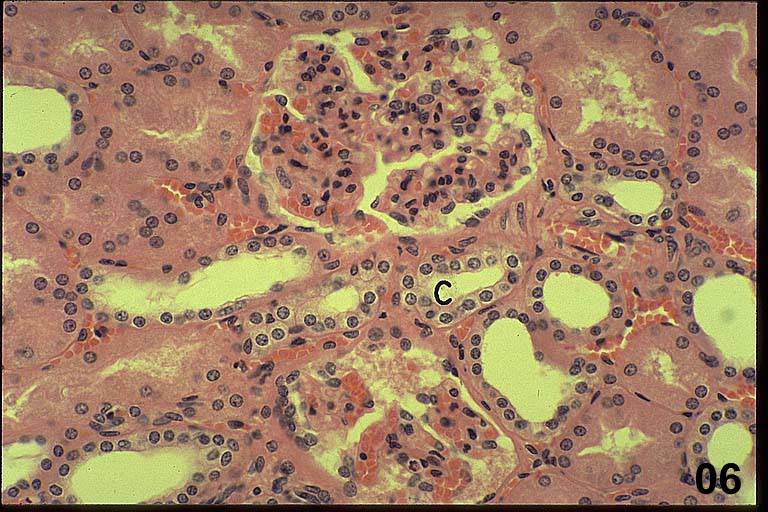
Within the cortex, distal convoluted tubules form arched collecting tubules (ACT) (HA6, C) that arch through the cortex and enter the medullary ray where they become collecting tubules. The cells of the arched collecting tubule have minimal lateral interdigitations so their lateral margins are visible. The cells have pale cytoplasm and their apical surfaces are often domed.
Key features of cortical tubules:
PCT - occluded lumen, no discernible lateral borders
DCT - obvious lumen, no discernible lateral borders, irregularly spaced nuclei
ACT – distinct lateral borders, domed cells
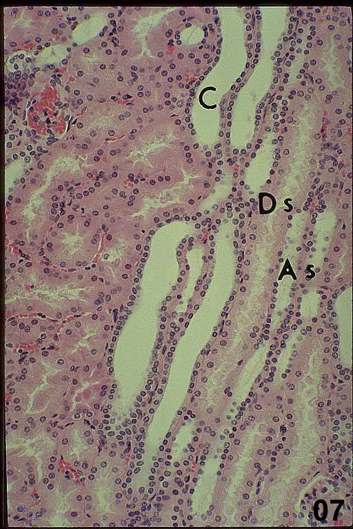
In the medullary ray there are parallel arrays of tubules shown here in longitudinal section (HA7). Note the similarities in morphology between the PCT in the cortex (left) and the descending thick limb (DTL) in the medullary ray (HA7, Ds). Both have irregularly shaped cells and occluded lumens. By comparison, the ascending thick limb (ATL) (HA7, As) has a larger lumen and shorter, more irregularly spaced nuclei than the PCT and DTL. The cells of the collecting tubule (CT) vary in height. If you are near the capsule, the collecting tubule cells are cuboidal (HA7, C). They become columnar as the tubule approaches and enters the medulla. The cells of the collecting tubule have obvious lateral cell borders and domed apices.
Key features of medullary ray tubules:
DTL - occluded lumen, no discernible lateral margins,
ATL - open lumen, no discernible lateral margins, irregularly spaced nuclei
CT - open lumen, domed apical surfaces, distinct lateral margins
All of the tubules that are found in the medullary ray can also be seen in the renal pyramid. The only difference is one name change: by definition, the collecting tubules become collecting ducts in the renal pyramid. The boundary is the arcuate arteries that define the corticomedullary junction.
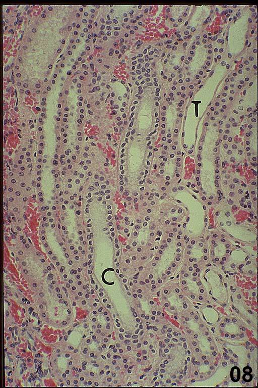
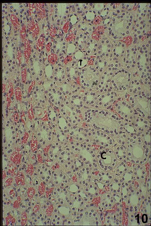
In the medulla that is close to the cortex ascending and descending thick limbs, thin limbs of Henle’s loop (HA8, T) and collecting ducts (HA8, C) are present. In the inner medulla, close to the papilla, only thin limbs (HA10, T) and collecting ducts (HA10, C) remain. As you move toward the apex of the renal pyramid, the epithelial cells of the collecting ducts get taller.
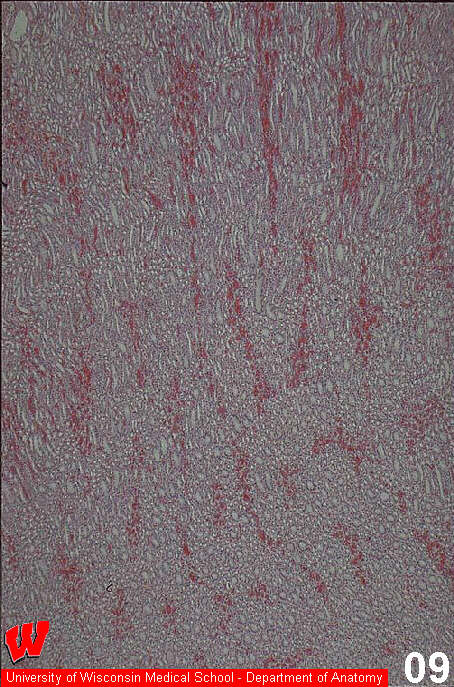
At low magnification, the overall arrangement of the vasae rectae (HA9), the straight vessels, can be seen. Because blood cells are still present in these vessels they are obvious even at low magnification and are easy to distinguish from the thin limbs of Henle’s loop (HA10, T) and the collecting ducts (HA10, C).
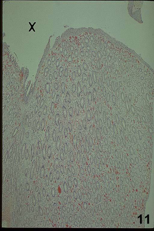
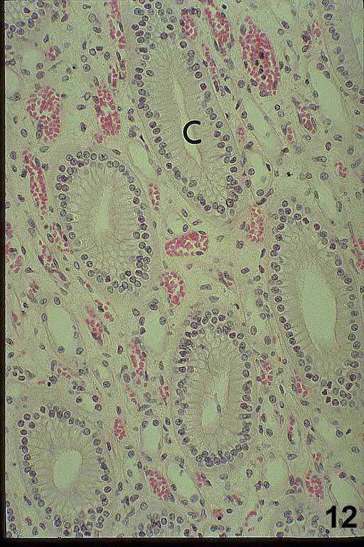
At the tip of the pyramid, the collecting ducts penetrate the urothelium covering the papilla and deliver urine into a minor calyx (HA11, X). The thin limbs, vasae rectae and collecting ducts (HA12, C) are all present in the papilla.