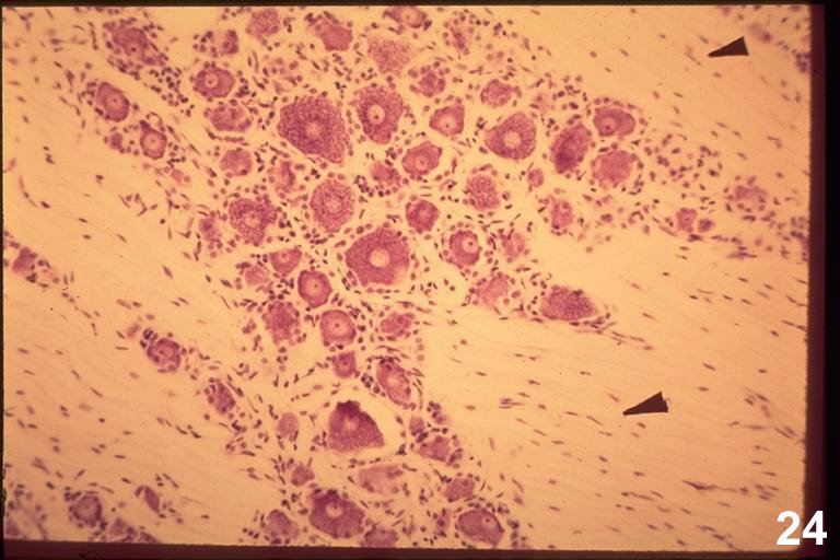
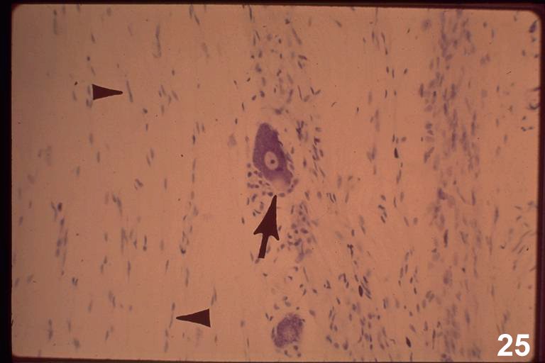
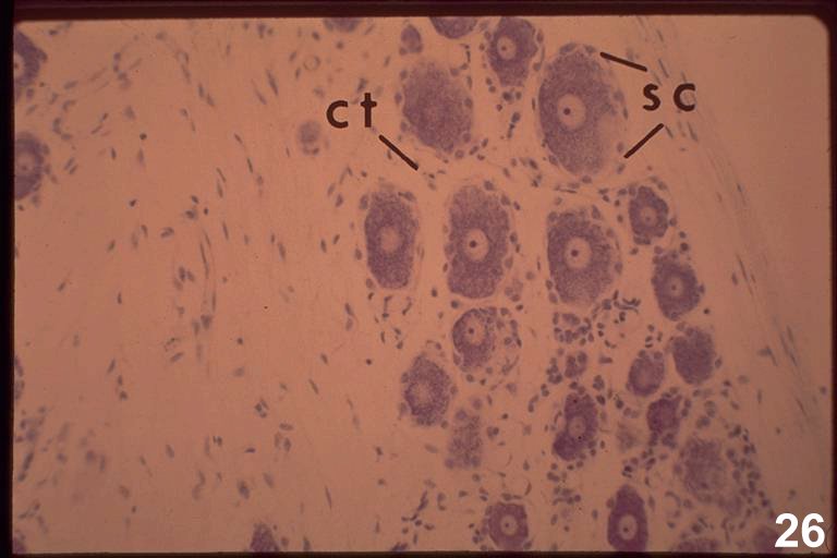
HA11-13: Dorsal root ganglion - Nissl stain
|
|
|
This image is of a longitudinal section of a dorsal root ganglion. The sensory neurons occur in narrow islands around which flow dorsal root axons ( arrowheads : HA11, 12 ).
Most of the nuclei that can be seen among the axons are those of Schwann cells. Some of the nuclei are those of endoneurial fibroblasts. It is impossible to tell which are fibroblast nuclei and which are Schwann cell nuclei.
The cell bodies of many of these neurons are large like those of the ventral motor horn cells and their contents are comparable. However, there are fundamental differences that are not visible in the light microscope. First, there are no dendrites and no synapses on the cell bodies. The dendrites of these neurons are out in the periphery at the ends of their axons. Second, the sensory neurons are unipolar. The axon has two components, a peripheral portion that is continuous with the dendrite of a receptor, and a central portion that runs into the CNS via the dorsal root.
It is easy to see the axon hillock (arrow : HA12 ). It is located at the perimeter of the cell body and can be identified by its lack of Nissl substance. In the EM, the hillock is seen to be packed with neurotubules and neurofilaments.
Satellite cells ( sc : HA13 ) have the same relation to PNS neuron cell bodies that Schwann cells have to PNS axons. They are interposed between the neuron and the endoneurial connective tissue. In this preparation, the small satellite cells appear to indent the neuron cell body. Their cytoplasm does not stain, so their nuclei are separated from the neuron cytoplasm by a thin, clear rim. Cells with flattened nuclei are also apparent that lie outside the satellite cells, between the neuronal soma. These are either endoneurial fibroblasts or Schwann cells ( ct: HA13 ). The perineurium and epineurium, surrounding the ganglion, are seen better in other preparations.