HA4-8: Ileum - fast green, hematoxylin & PAS
|
|
|
|
|
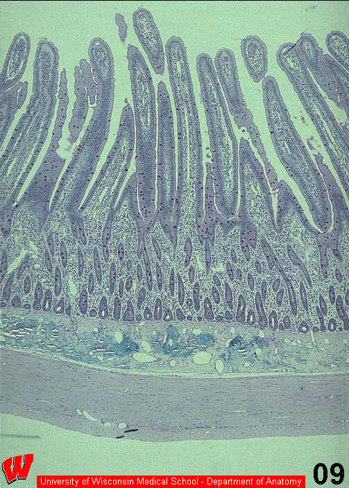
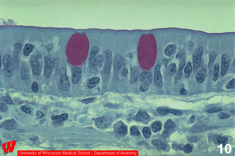
Refer to the diagram in your coursebook to begin to understand the 3-dimensional structure of the lining of the small intestine. Begin by looking at the lumen of the ileum (top, HA4 ). Finger-shaped structures, called villi project into the lumen. The epithelium covering the villi has many different appearances, but the thinnest area is simple columnar epithelium, as shown in this high magnification image (HA5) which is perpendicular to HA4 . Careful examination of this light micrograph reveals two stripes at the apical surface (top), one pink stripe and one green stripe (HA5) . The pink stripe is the glycocalyx, a layer of sugars linked to the plasmalemma that stains pink with the sugar-specific PAS stain. The green stripe is the microvillous border. At the base of the microvilli there is a thin darker green line, the terminal web. The terminal web inserts at every intercellular boundary into a slight thickening called the terminal bar, where a junctional complex (see below) exists between the cells. Scattered throughout the epithelium, there are magenta ovals, which represent the apical cytoplasm of goblet cells, unicellular exocrine glands that secrete mucus onto the apical surface. The complete name of this epithelium is simple columnar epithelium with microvilli and goblet cells. This type of epithelium is found throughout the small intestines. What is the function of the microvilli on this epithelium?
Note that the villus is a gross anatomical structure easily seen at low magnification, where as microvilli are difficult to see even at high magnification in a light micrograph.
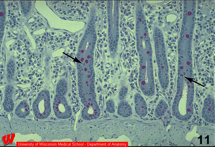
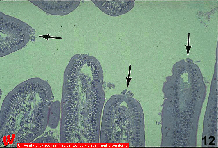
The image in HA6 is deep in the wall of the ileum and shows many crypts cut in different planes of section surrounded by a CT layer called the lamina propria. Cells in the crypt divide ( HA6, arrows ) and "push" the epithelium toward the tip of the villus in the process of cell turnover (HA6) . The mitotic cells in this part of the epithelium provide a constant supply of new cells, while other cells are being sloughed into the lumen at the tip of the villus (HA7, arrows) . This epithelium is replaced completely every 5-7 days. What are the darkly staining structures in the mitotic cells at the tips of the arrows in HA6 ?
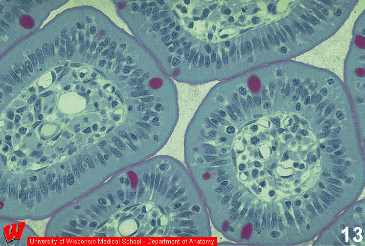
The micrograph in HA8 shows several villi cut in cross-section. The CT layer called lamina propria lies at the center of each villus. The lumens are either blood or lymph vessels. As described for HA5 , the epithelium is simple columnar, but in many places it appears stratified. This is not due to plane of section. It is due to the small lymphocytes that are in between the epithelial cells. The lymphocytes have a halo-like cytoplasm and dark nuclei, and oten do not touch either the apex or the base of the epithelium. When naming epithelia shown in a micrograph, always look for the thinnest section through an epithelium to name it. Why do you think it is important to have immune system cells so close to the lumen of the intestine?